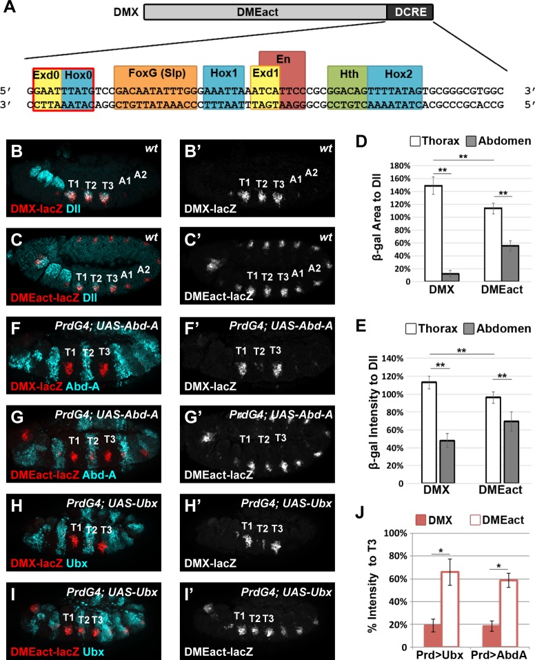
Fig 1. The abdominal Hox factors repress Distal-less via DCRE-dependent and–independent mechanisms.
(A) Schematic of the DMX enhancer containing the DMEact and the DCRE. Detail shows the DCRE sequence with known TF binding sites (FoxG, Hox1, Exd, En, Hth, and Hox2) highlighted. Note, the Exd0 and Hox0 sites are new sites characterized in this manuscript. (B) DMX-lacZ is expressed in Dll-positive (Dll+) cells of the thorax. (C) DMEact-lacZ is expressed in Dll+ cells of the thorax and the corresponding cells of the abdomen. (D-E) Quantification of β-gal expression area (D) and β-gal intensity relative to Dll expression in the same embryos (E) in DMX-lacZ and DMEact-lacZ embryos demonstrates that the DMEact is not fully de-repressed in the abdomen and has reduced thoracic levels when compared to thoracic levels of DMX. (F-I) Effect of Hox gene mis-expression on reporter activity in PrdG4;UAS-AbdA;DMX-lacZ (F), PrdG4;UAS-AbdA;DMEact-lacZ (G), PrdG4;UAS-Ubx;DMX-lacZ (H), and PrdG4;UAS-Ubx;DMEact-lacZ (I) embryos. (J) Quantification of β-gal levels in T2 mis-expressing segments relative to non-Gal4 expressing control T3 segments shows that both Abd-A and Ubx repress via DCRE-dependent and -independent mechanisms. All images are lateral views of Stage 11 embryos immunostained for β-gal (red or white) and Dll, Abd-A, or Ubx (cyan) as indicated. (Statistics ** p < 0.01, Welch’s t-test, error bars S.E.M). Note, as a control for the PrdG4 experiments, we quantified the levels of βgal in the absence of Gal4 and noted no significant differences between the T2 vs T3 segments or the A1 vs A2 segments of DMX-lacZ and DMEact-lacZ embryos. (DMX β-gal pixel intensity: Thorax, T2 = 113 ± 13%, T3 = 112 ± 10%, Abdomen, A1 = 44 ±10%, A2 = 56 ± 13%. DMEact β-gal pixel intensity: Thorax, T2 = 98 ± 12%, T3 = 96 ± 11, Abdomen, A1 = 69 ±12%, A2 = 70 ± 16%).

