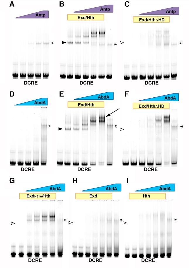
Fig 6. Hox binding to linked Hox/Exd and Hox/Hth sites within the DCRE.
(A-I) EMSAs performed on the DCRE full-length probe (for sequence, see Fig 1A). (A-C) Titration of Antp protein (concentrations from 37.5 nM to 300 nM) alone (A), with 25 nM purified Exd/Hth dimer (B), or with 200 nM purified Exd/HthΔHD dimer (C). (D-I) Titration of AbdA protein (concentrations from 37.5 nM to 300 nM) on the DCRE wild-type probe alone or with (D) 75 nM purified Exd/Hth dimer (E) 200 nM purified Exd/HthΔHD dimer (F), 200nM purified ExdN51A/Hth (G), 200 nM purified Exd (H), or 200 nM purified Hth (I). * indicates Hox-only binding to DCRE. Arrow indicates higher order complex seen with AbdA/Exd/Hth but not Antp/Exd/Hth. Filled arrowheads indicate Hth/Exd heterodimer binding. Empty arrowheads indicate lack of binding by Hth and/or Exd.

