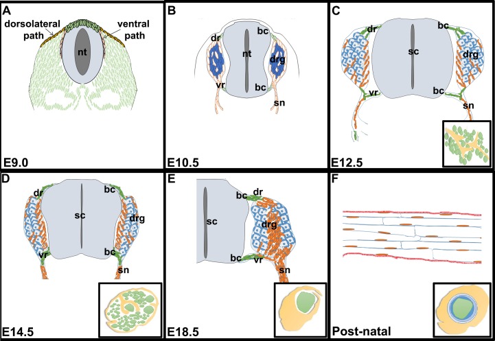
Fig 1. Schematic representation of the different phases of Schwann cell development.
Schematic images of transverse sections through the trunk of E9.0 (A), E10.5 (B), E12.5 (C), E14.5 (D) and E18.5 (E) embryos, and a longitudinal section through the postnatal sciatic nerve (F). Insets in C-F show transverse sections through the sciatic nerve. bc, boundary cap; dr, dorsal root; drg, dorsal root ganglia; nt, neural tube; sn, spinal nerve; vr, ventral root.

