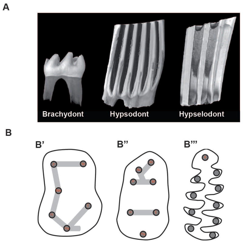
Figure 1.

Schematic representation of dental morphological characters. (A) 3D renderings obtained by X-ray microtomography illustrating three different levels of lower first molar crown height. From left to right: brachydont (Mus musculus), hypsodont (Ondatra zibethicus), and euhypsodont (Lemmus lemmus). (B) Cartoons of occlusal surface organization. From left to right: lower first molar occlusal surface of basal forms, murine rodents (B′ and B″) (including Mus musculus), and arvicoline rodents (B‴) (including Microtus californicus). Cusp naming abbreviations (in red): P, Protoconid: M, Metaconid; Me, Mesoconid; H, Hypoconid; E, Entoconid: Hy, Hypoconulid; A, Anteroconid; Av, Vestibular anteroconid; Al, Lingual anteroconid. 3D-renderings and cartoons not at scale to facilitate morphological comparisons. Whereas rounded cusps connected by lophs characterize the murine phenotype, triangular cusps along a main ridge characterize the arvicolin phenotype.
