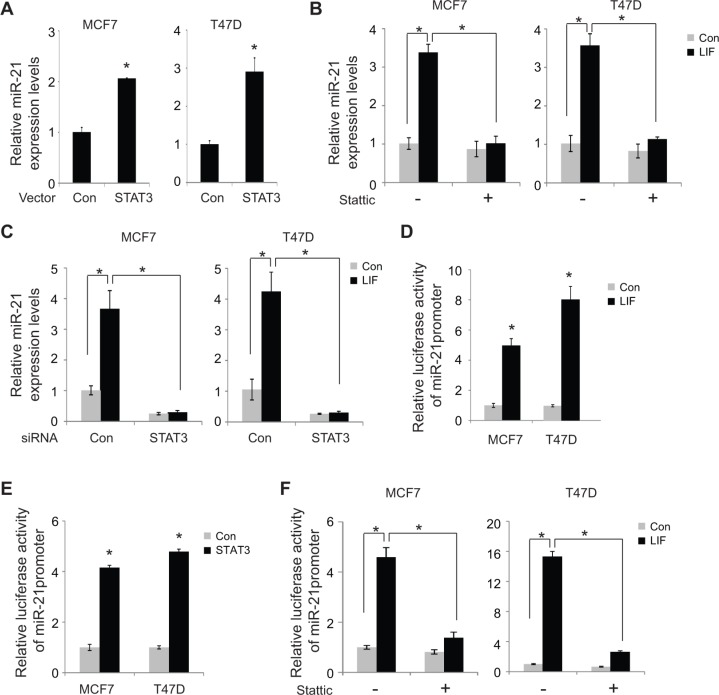
Figure 6. LIF induces miR-21 expression through STAT3.
(A) MCF7 and T47D cells were transfected with STAT3 expression vectors or control vectors. The expression levels of miR-21 were determined at 24 h after transfection by real-time PCR. The expression of miR-21 was normalized to the U6 snRNA. (B) Stattic treatment largely abolished the induction of miR-21 by LIF. MCF7 and T47D cells with stable ectopic LIF expression and their control cells were treated with Stattic (2 μM), and the miR-21 expression levels were determined by real-time PCR. (C) Knockdown of endogenous STAT3 by siRNA oligos largely abolished the induction of miR-21 by LIF. MCF7 and T47D cells with stable ectopic expression of LIF and their control cells were transfected with siRNA oligos targeting STAT3 or control siRNA. The expression levels of miR-21 were determined using real-time PCR. (D) MCF7 and T47D cells with or without ectopic LIF expression were transfected with the luciferase reporter vectors containing the miR-21 promoter together with pRL-null vectors as an internal control to normalize transfection followed by luciferase activity measurement. The luciferase activities of reporter vectors were normalized to internal control. (E) MCF7 and T47D cells were transfected with the luciferase reporter vectors containing the miR-21 promoter together with pRL-null vectors and STAT3 expression vectors. (F) MCF7 and T47D cells transfected with luciferase reporter vectors together with LIF expression vectors or control vectors were treated with Stattic (2 μM). Luciferase activities were measured after 24 h of treatment. Data are presented as mean ± s.d. (n = 3). *p < 0.05, student t-test.

