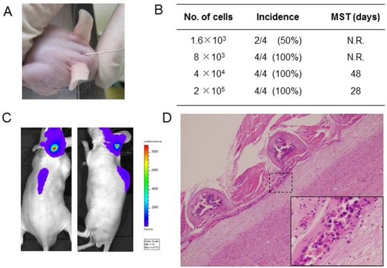
Figure 2. Leptomeningeal carcinomatosis model with PC-9/ffluc cells.
(A) PC-9/ffluc cells were inoculated into the space between the external occipital protuberance and first cervical vertebra of SHO-SCID mice. (B) Production of LMC was determined using the IVIS imaging system; MST: median survival time; N.R: not reached. (C) Representative imaging of mice immediately after PC-9/ffluc cell inoculation. (D) Representative histological images of spinal cord with LMC (x40). The high-power field image of dotted area is shown in the lower right corner (x400).

