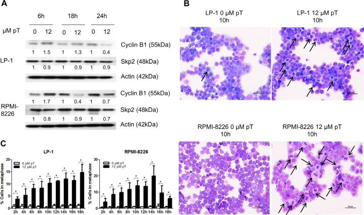
Figure 2. Pharmacological inhibition of the APC/C with proTAME results in a metaphase arrest.
(A) LP-1 and RPMI-8226 cells were treated with 12 μM proTAME (pT) for 6, 18 and 24 hours. Western blot analysis was performed using cyclin B1, Skp2 and β-actin antibodies. This result is representative for 3 independent experiments. The pixel densities of proteins are normalized to β-actin and relative to control. (B) LP-1 and RPMI-8226 cells were treated with 12 μM proTAME and every 2 hours until 18 hours May-Grünwald Giemsa stained cytospins were made. Arrows point out the MM cells in metaphase. Scale bar measures 50 μm. (C) Quantification of the amount of cells in metaphase. Results shown in each graph are the mean of 3 independent experiments ± SD. *means the p-value is < 0.05 (Mann-Whitney U-test).

