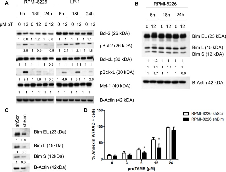
Figure 6. proTAME treatment induces Bim and phosphorylation of Bcl-2 and Bcl-xL.
(A and B) LP-1 and RPMI-8226 cells were treated with 12 μM proTAME for 6, 18 and 24 hours. Western blot was performed using Bcl-2, pBcl-2, Bcl-xL, pBcl-xL, Mcl-1, Bim and β-actin antibodies. The pixel densities of proteins were normalized to β-actin and relative to control. This result is representative for 3 independent experiments. (C) Bim was knocked down in the RPMI-8226 using shRNA. These cells were treated with 0, 3, 6, 12 and 24 μM proTAME and after 48 hours apoptosis was measured with Annexin V/7AAD flow cytometry staining. Results shown in each graph are the mean of 4 independent experiments ± SD,*means the p-value is < 0.05 and indicates a significant difference compared to RPMI-8226shScr (Mann-Whitney U-test) (D).

