
Figure 3. Reduced cell growth after the VDAC1 gene had been silenced in SiHa and CaSki cervical cancer cells.
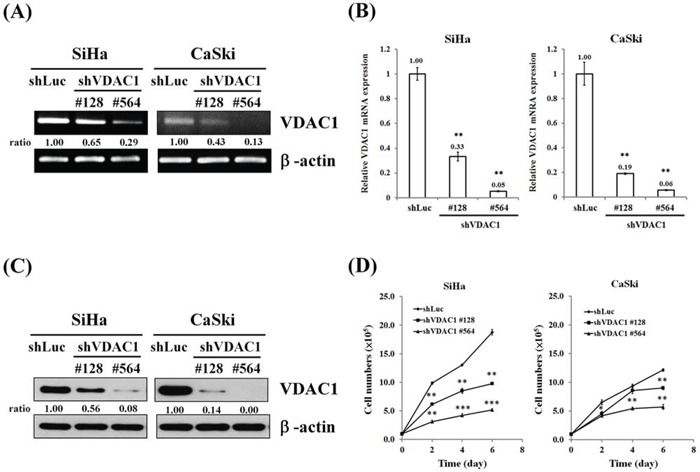
SiHa and CaSki cells (5×10⁵ cells/6-cm dish) were infected with lentiviruses carrying shVDAC1 #128, #564 or vector control (shLuc). The RNA levels were determined by A. RT-PCR and B. real-time PCR. β-actin was used as the internal control. The relative ratios of VDAC1/β-actin are shown. C. Western blotting was used to detect the VDAC1 expression in SiHa or CaSki shVDAC1 #128, #564 and shLuc cells. β-actin was used as the internal control. The relative ratios of VDAC1/β-actin are shown. D. SiHa or CaSki shVDAC1 #128, #564 or shLuc cells (1 × 10⁵ cells/6-cm dish) were seeded and then analyzed for growth curves for 2, 4 and 6 days by counting cell numbers. All values are means ± SD from at least three independent experiments. *p<0.05, **p<0.01, ***p<0.001.