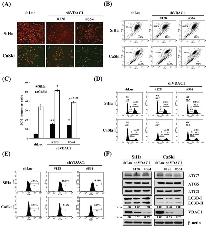
Figure 4. The effects of shVDAC1 on MMP, cell cycle, ROS production and autophagy.

A. Representative images of SiHa and CaSki shVDAC1 #128, #564 and shLuc cells stained with JC-1 (magnification, ×100). White arrows indicate green fluorescence (monomeric JC-1). B. MMP measurements by flow cytometry and the intensity of red fluorescence (R1) and green fluorescence (R2) of JC-1. C. Bar graph shows changes in quantification in JC-1 fluorescence as detected by flow cytometry assay. The JC-1 monomer ratio was increased in VDAC1 gene-silenced SiHa or CaSki cervical cancer cells compared to their control counterparts. Data are presented as mean ± SD. *p<0.05, **p<0.01. D. The cell cycle of SiHa and CaSki shVDAC1 #128, #564 or shLuc cells was analyzed by propidium iodide staining using flow cytometry. No significant changes were found in the VDAC1 gene-silenced cancer cells. E. Increased ROS generation was found in the VDAC1 gene-silenced cervical cancer cells, and especially in SiHa shVDAC1 #128 and #564 cancer cells. The cells were stained with 10 μM H2DCFDA for 30 minutes. ROS production was measured by flow cytometry. F. The protein levels were determined by Western blotting. β-actin was used as the internal control. The relative ratios of LC3B-II/β-actin and VDAC1/β-actin are shown. The VDAC1 gene was silenced using lentiviruses carrying shVDAC1 #128 and #564 in SiHa and CaSki cervical cancer cells. Control vector: shLuc. VDAC1, voltage-dependent anion channel 1; MMP, mitochondrial membrane potential; ROS, reactive oxygen species; Atg, autophagy-correlated protein; LC3, microtubule-associated protein light chain 3.
