
Figure 5. Reduced cell migration in cervical cancer cells in which the VDAC1 gene had been silenced.
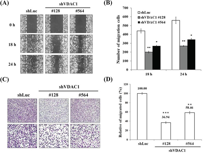
A. Wound healing arrays were performed on SiHa shVDAC1 #128, #564 and shLuc cells (2 × 10⁴ cells/well of culture inserts). Images were captured at the indicated times after the initial wound in the top panels (magnification, ×100). B. Cell motility was significantly reduced after the VDAC1 gene had been silenced in SiHa cervical cancer cells. The number of cells that had migrated were counted under a microscope. C. Reduced cell migration using a Boyden chamber with a polycarbonate membrane (12-μm pore size) after the VDAC1 gene had been silenced in SiHa cervical cancer cells. A representative microscopic image for each condition is shown (magnification, ×40 and ×100). D. Cell migration was significantly reduced after the VDAC1 gene had been silenced in SiHa cervical cancer cells. The migration rate was determined by counting the cells that had migrated through the polycarbonate membrane and was expressed as the relative percentage to those with the control vector (shLuc, set to 100%). All data are representative of at least three different experiments. Values are expressed as mean±SD. The VDAC1 gene was silenced using lentiviruses carrying shVDAC1 #128 and #564 in SiHa cervical cancer cells. VDAC1, voltage-dependent anion channel 1. *p<0.05, **p<0.01, ***p<0.001.