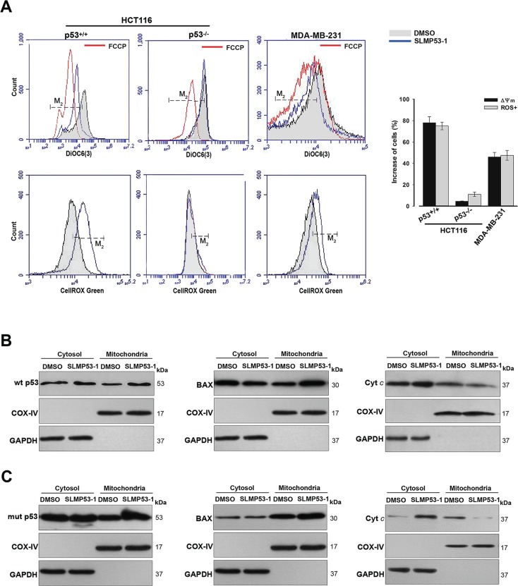
Figure 4. SLMP53-1 triggers a p53-mediated mitochondrial apoptotic pathway in HCT116p53+/+ and MDA-MB-231 cells.
(A) Cells were treated with 16 μM SLMP53-1 for 8 h (HCT116 cells) or 16 h (MDA-MB-231 cells) in Δψm analysis, and for 24 h in ROS analysis. Carbonyl cyanide-p-trifluoromethoxyphenylhydrazone (FCCP) was used as positive control in Δψm; histograms are representative of three independent experiments; M2 cursor indicates the subpopulation analyzed. Graphical representation: increase in the percentage of cells with Δψm dissipation and ROS generation; values are mean ± SEM (n = 3). (B) and (C) Immunoblots of wt/mut p53, BAX and cyt c, in cytosolic and mitochondrial fractions of (B) HCT116p53+/+ and (C) MDA-MB-231 cells after 16 h with 16 μM SLMP53-1. In B and C, immunoblots are representative of three independent experiments.

