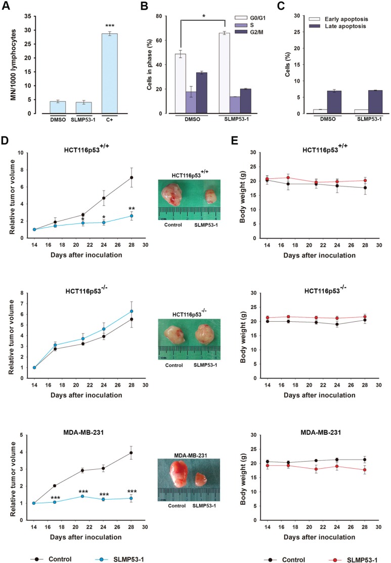
Figure 7. SLMP53-1, with no apparent in vitro toxicity, has potent in vivo antitumor activity.
(A) Genotoxicity of 16 μM SLMP53-1 by cytokinesis-block micronucleus (MN) assay after 72 h treatment in human lymphocyte cells. Cells treated with 1 μg/mL cyclophosphamide were used as positive control (C+); data are mean ± SEM (n = 3). (B) Cell cycle after 48 h with 42.4 μM SLMP53-1 in MCF10A cells; data are mean ± SEM (n = 3). In A and B, values significantly different from DMSO only: ***p < 0.001. C. Apoptosis after 48 h with 42.4 μM SLMP53-1 in MCF10A cells; data are mean ± SEM (n = 3); no significant differences between SLMP53-1 and DMSO only: p > 0.05. (D) BALB/c nude mice carrying HCT116p53+/+, HCT116p53−/− or MDA-MB-231 xenografts treated with 50 mg/kg SLMP53-1 or vehicle (control); values significantly different from control mice: *p < 0.05, **p < 0.01, ***p < 0.001; representative pictures of control and SLMP53-1-treated tumors. (E) BALB/c mice body weight during SLMP53-1 treatment; no significant differences between control and SLMP53-1-treated mice: p > 0.05.

