
Figure 6. Genetic inhibition of autophagy promotes TRAIL-induced cell death upon niacin treatment.
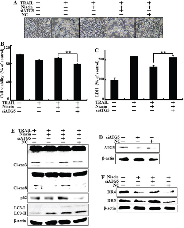
HCT116 cells were pre-treated with 20 nM ATG5 siRNA for 4 h and then exposed to 800 μM niacin for 12 h and treated with 100 ng/ml TRAIL for 2 h. A. Cell morphology was photographed under light microscopy (×200). B. Viability of treated cells was measured by crystal violet staining. Viability of control cells was taken as 100%. C. LDH release into the cell culture medium was measured after exposure to TRAIL for 2 h. D. Western blot analysis of ATG5 protein confirmed specific protein knockdown. β-actin was used as a loading control. E. Western blot analysis of caspase-3 and caspase-8. β-actin was used as a loading control. F. Western blot analysis data showed DR4 and DR5 protein known as TRAIL related death receptor. β-actin was used as a loading control. *p < 0.05 or **p < 0.01 significant differences between control and each treatment group.