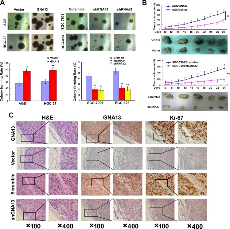
Figure 4. GNA13 promotes the tumorigenicity of GC cells in vitro and in vivo.
(A) Anchorage-independent growth assay in GNA13-overexpressing cells (Left) and GNA13-silenced cells (Right). Soft agar colony formation (colonies larger than 0.1 mm diameter) was quantified after 14 days of culture (Lower panel). (B) AGS/GNA13 and AGS/Vector cells, and SGC-7901/shGNA13 and SGC-7901/scramble cells were injected in the hindlimbs of nude mice (n = 5). Tumor volumes were measured on the indicated days. (C) Histopathology of xenograft tumors. The tumor sections were under H&E staining and IHC staining using antibodies against GNA13 and Ki-67. *P < 0.05; **P < 0.01.

