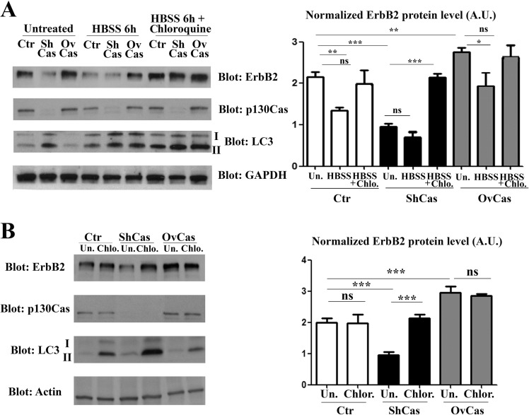
Figure 3. p130Cas protects ErbB2 from autophagy-dependent degradation.
(A) Left panel: Extracts from p130Cas silenced, overexpressing and control (Ctr) BT474 cells cultured for 6 hours in HBSS in absence or presence of chloroquine (100 μM) were blotted with antibodies to ErbB2, p130Cas, LC3 and GAPDH as loading control. Right panel: Histograms show ErbB2 levels, normalized to GAPDH. Bars represent the means ± SEM of five independent experiments (ns: not significant; **p < 0.01; ***p < 0.001). (B) Left panel: BT474 cells as in (A) were treated with 100 μM chloroquine for 6 hours. Cell lysates were blotted with antibodies against ErbB2, p130Cas, LC3 and Actin as loading control. Right panel: Histograms show ErbB2 levels, normalized to Actin. Bars represent the means ± SEM of four independent experiments (ns: not significant; ***p < 0.001).

