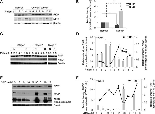
Figure 2. RKIP and NICD are inversely expressed in human cervical and stomach cancer tissues.

(A, B) RKIP and NICD expression in human cervical cancer. (A) Proteins were extracted from normal (patients #1–3) and cancerous (patients #4–11) human cervical tissues. Total proteins (30 μg) were subjected to immunoblot analysis. (B) Expression of each protein (RKIP or NICD) was quantified by densitometric scans and represented graphically. *p < 0.05 compared to normalized controls. (C, D) RKIP and NICD expression in human stomach cancer. (C) Proteins were extracted from pairs of tumorous and adjacent non-tumorous tissues of nine human stomach cancer patients (patients #1–9) with three different TNM stages (stage 1: patients #1–4, stage 2: patients #5–7, and stage 3: patients #8–9). (D) RKIP and NICD expression in stomach cancer specimens (indicated by “C”) were quantified followed by normalization to the expression of each protein, respectively, in matched adjacent non-tumorous tissues (indicated by “N”) and represented graphically. *p < 0.05 compared to normalized controls. (E, F) Expression of RKIP and NICD in YCC gastric cancer clones (human advanced gastric cancer cell lines). (E) Nine YCC clones (#3, 6, 7, 9, 10, 16, 18, 21, and 36) were subjected to immunoblot analyses. (F) The expression levels of RKIP and NICD in YCC clones were quantified and represented graphically. β-actin was used as loading control. All data indicate the mean values ± S.D. of at least three independent experiments. *p < 0.05.
