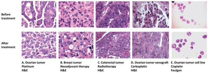
Figure 1. Nuclear morphology changes in different clinical and experimental settings.

Similar nuclear texture changes occur in: A. ovarian tumors after chemotherapy; B. breast tumors after neoadjuvant therapy; C. colorectal tumors after radiotherapy; D. ovarian tumor xenografts after carboplatin treatment; and E. the ovarian cancer cell line PEO1 after cisplatin treatment. A-D are formalin-fixed, paraffin embedded sections cut from tumor samples and stained with H&E. E shows PEO1 cell cytospins using the Feulgen nuclear stain.
