
Figure 3. Changes in apoptosis and necroptosis of leukemia cells after lentiviral infection and VP16 treatment (48 h).
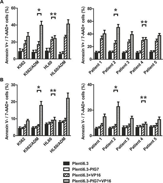
Annexin V staining assay indicated that (A) the apoptosis rates of these four cell lines and five cases of primary cells infected with Plent6.3-PIG7 lentivirus were not changed. When combined with differential concentrations of VP16, the apoptotic rate of these three cell lines and four cases of primary cells significantly increased when compared with Plent6.3 control + VP16 groups. K562/ADM and Patient 2 has the highest increase in apoptosis rate (*). There was almost no change in apoptosis rate in either HL60 or Patient 4 cells (**P > 0.05) (B) The necroptosis rate of cells infected with Plent6.3-PIG7 lentivirus was unchanged. When combined with VP16, the necroptosis rate of all cells increased compared with Plent6.3 control + VP16 groups. K562/ADM and Patient 2 had the highest increases in their respective necroptosis rates (*), while HL60 and Patient 4 had the lowest increases in their necroptosis rates (**).