
Figure 4. Overexpression of pig7 induces LMP.
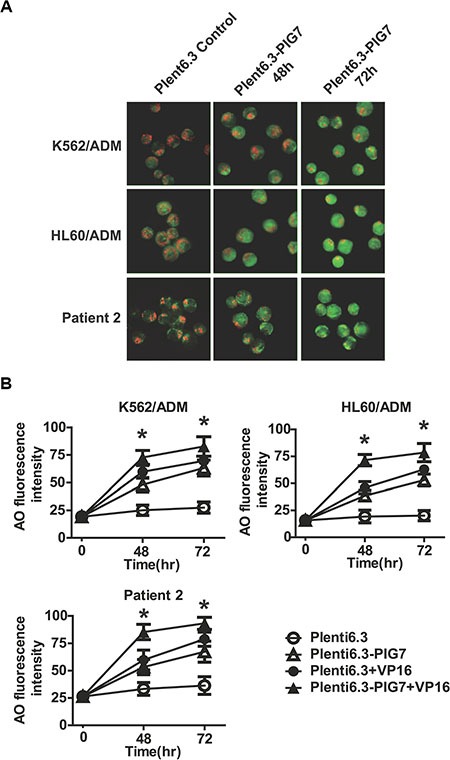
(A) LMP detected by AO staining and visualized by fluorescence microscopy. (B) Percentage of leukemia cells displaying LMP quantified by flow cytometric analysis of AO staining after lentiviral infection in combination with VP16. 48 and 72 h green fluorescence intensities both significantly increased in the Plent6.3-PIG7 group. The Plent6.3-PIG7 + VP16 group had the highest green fluorescence intensity (*P < 0.05).