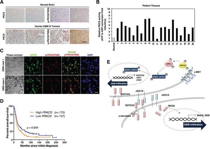
Figure 5. Clinical relevance of PKCδ in GBM patients.

A. Immunohistochemistry for PKCδ and p-PKCδ in human normal brain tissues and GBM patients (n = 30). Scale bar, 200 μm. B. PKCδ kinase activity in normal brain tissue and 20 cases of human GBM patients. C. Immunohistochemistry for co-staining of PKCδ and NICD2 in human GBM. Scale bar, 100 μm. D. Kaplan-Meier survival curves of high and low levels of PKCδ in human brain tumor patients with REMBRANDT database. E. Schematic model illustrating the PKCδ-associated signaling pathways leading to infiltration of GBM.
