
Fig. 1.
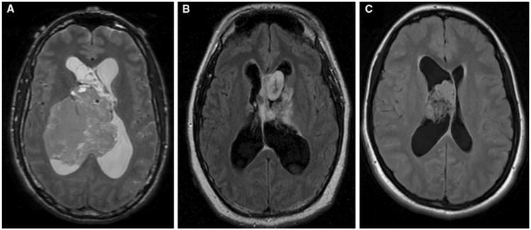
Representative neurocytoma MRI imaging findings. a T2 weighted imaging of a 44-year-old male found to have an 8.6 × 7.2 cm heterogeneous right lateral ventricle mass; predominantly isointense on T2 weighted imaging with no significant enhancement on post-contrast series. b T2 FLAIR imaging of a 23-year-old male found to have a heterogeneous lobulated mass with a mildly enhancing component and other non-enhancing components extending superiorly from the suprasellar cistern. c T2 FLAIR imaging of a 20-year-old female found to have a 3.4 × 2.5 cm highly cellular lobulated nonenhancing intraventricular mass attached to the anterior portion of the septum pellucidum