Fig. 2.

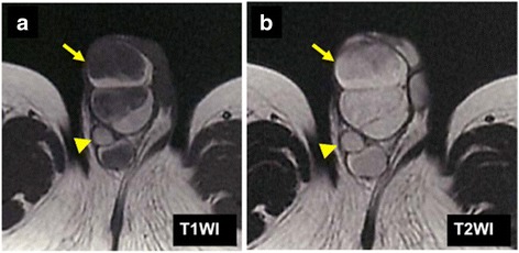
The MRI findings. a T1-weighted image showed the mosaic pattern (e.g., T1-low lesions and T1-high lesions were co-existed). b All of the tumor revealed the high intensity on T2-weighted image. (Arrow heads indicate right testis.)

The MRI findings. a T1-weighted image showed the mosaic pattern (e.g., T1-low lesions and T1-high lesions were co-existed). b All of the tumor revealed the high intensity on T2-weighted image. (Arrow heads indicate right testis.)