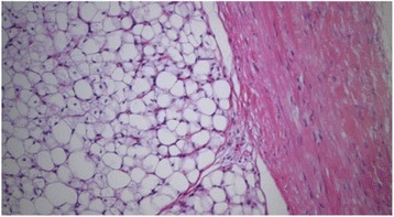
Fig. 5.

Pathological analysis. Microscopically, both tumors were multilobulated tumors of adipose tissue and were surrounded by the capsule composed of loose connective tissue. The both tumors were diagnosed as lipoblastoma (hematoxylin-eosin stain, ×100)
