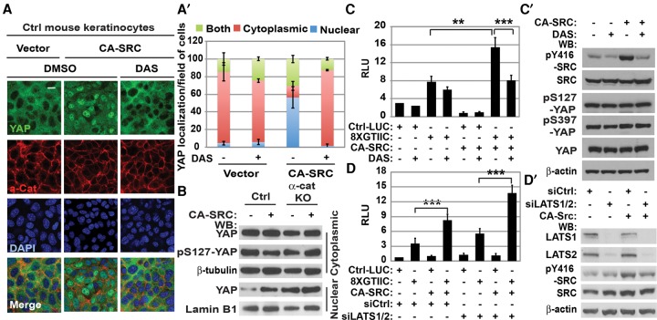
Figure 3.
Active SRC promotes Hippo pathway-independent nuclear localization and activation of YAP1 in confluent keratinocytes. (A) Immunofluorescent staining of keratinocytes stably transduced with vector or CA-SRC and treated with DMSO or DAS with anti-αE-catenin (α-cat; red) and anti-YAP1 (green) antibodies. DAPI indicates nuclear counterstain (blue). Cells were treated with either vehicle (DMSO) or 5 nM DAS for 30 min at 37°C. Bar, 15 µm. (A′) Quantitation of YAP1 localization in A. Bars represent the average of YAP1 localization between the cytoplasm, the nucleus, or both ±SD for >150 cells for each condition. (B) Western blot (WB) analyses of proteins from nuclear and cytoplasmic fractions of control (Ctrl) or α-cat knockout (KO) mouse keratinocytes transduced with vector (−) or CA-SRC (+). (C) Luciferase assay using empty control (Ctrl-LUC) or TEAD/YAP1 8xGTIIC reporter in keratinocytes stably transduced with empty vector or CA-SRC. DAS indicated 0.2 nM DAS. Bars represent means ± SD. n = 3. Student's t-test. (C′) Western blot analyses of total proteins from keratinocytes expressing empty control or CA-SRC constructs and treated with DMSO (−) or DAS using the indicated antibodies. (D) Luciferase assay using control (Ctrl-LUC) or the TEAD/YAP1 8xGTIIC reporter in keratinocytes stably transduced with empty vector or CA-SRC and transiently transfected with siCtrl or siLATS1/2 oligos. Bars represent means ± SD. n = 3. Student's t-test. (D′) Western blot analyses of total proteins from keratinocytes expressing empty control or CA-SRC constructs and transiently transfected with siCtrl or siLATS1/2 oligos using the indicated antibodies.

