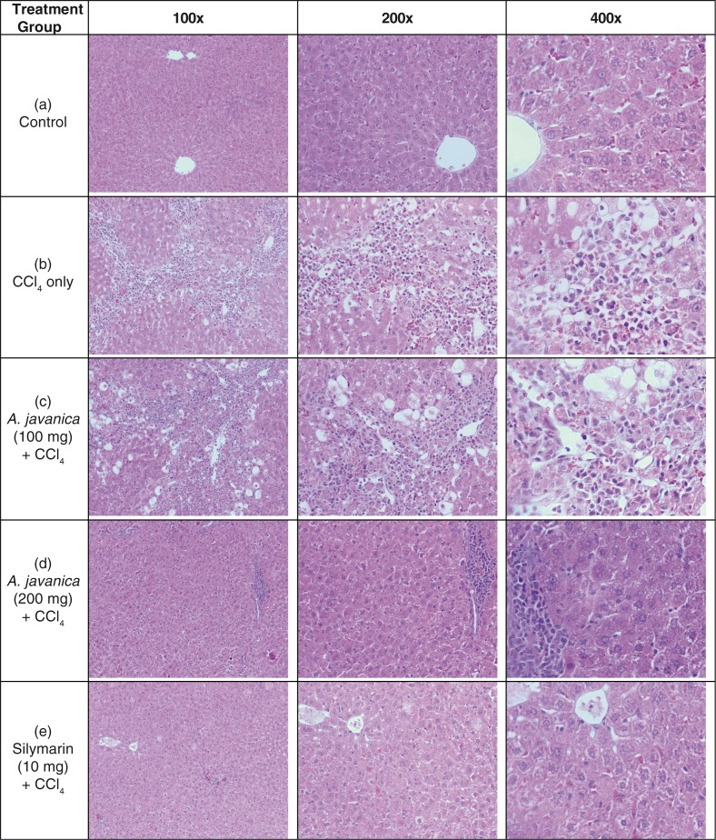
Fig. 2.
The histopathology of experimental rat liver at 100×, 200×, and 400× magnifications. Histograms showing the following: (a) healthy tissues with normal hepatocytes and central vein; (b) CCl4-injured tissue with necrosis and fatty degenerative changes; (c) tissue with congested central vein with necrosis and fatty changes after A. javanica (100 mg) + CCl4 treatment; (d) liver with normal hepatocytes and central vein with full recovery after A. javanica (200 mg) + CCl4 treatment; and (e) liver with normal hepatocytes and fully recovered central vein after silymarin (10 mg) + CCl4 treatment.

