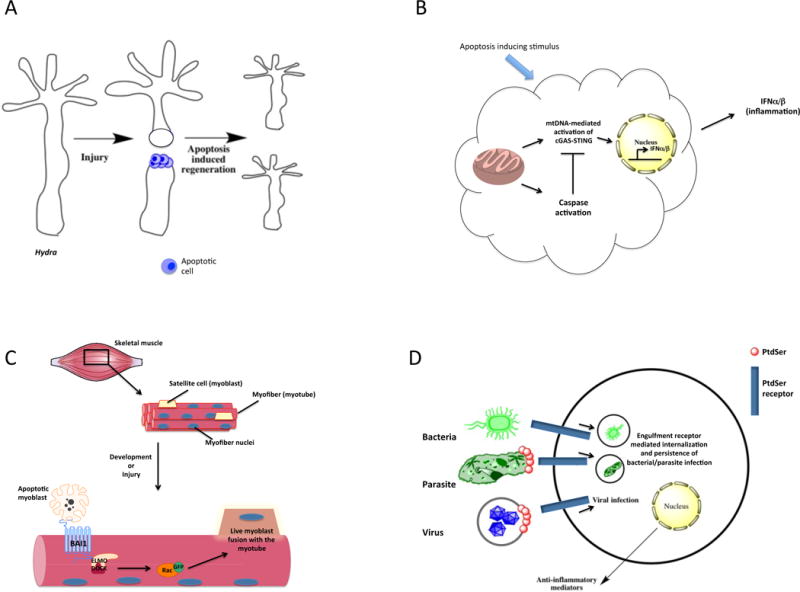
Fig. 4.

Additional and non-obvious functions of apoptotic cells. (a) Regeneration: In the metazoan Hydra, tissue injury can lead to apoptosis of the cells, which stimulates regenerative processes in the nearby viable tissues via a process called ‘apoptosis-induced compensatory proliferation’. Apoptosis is required for the re-growth of the new Hydra head. (b) Caspase-dependent inhibition of interferon production: In the context of a viral infection, apoptosis leads to the activation of caspases that is linked to inhibiting interferon-α/β (IFN-α/β) production induced by the mitochondrial DNA (mtDNA)-mediated activation of the cGAS-STING pathway. (c) Myoblast fusion: During muscle development and regeneration after muscle injury, apoptosis of myoblasts triggers BAI1 signaling through the ELMO-DOCK complex, leading to Rac activation. This pathway contributes to the fusion of healthy myoblasts with the nascent myotube and promotes muscle development and regeneration. (d) Pathogen exploitation of engulfment receptors: Bacteria, parasites and even viruses have evolved to utilize the apoptotic cell engulfment receptors for the cellular entry and induction of anti-inflammatory signaling in the host cell (phagocyte), aiding in the establishment and persistence of infection.
