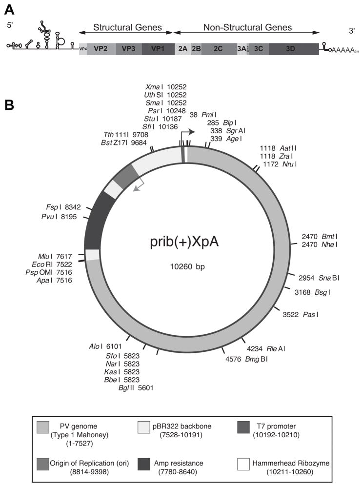
Figure 1.
(A) Schematic of the PV Genome. The PV genome includes a highly structured 5′UTR followed by a single open reading frame and a polyadenylated 3′UTR. The coding region can be separated into structural and non-structural genes. The structural genes are required to form the viral capsid and the non-structural genes are all required for a successful replication cycle. (B) prib(+)XpA plasmid map. PV1 (Mahoney) genome is in the pBR322 backbone. The plasmid contains an ampicillin resistance cassette, a T7 promoter, and a hammerhead ribozyme. Location of single cut restriction sites are shown.

