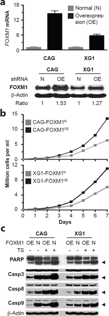
Figure 4. Enforced expression of FOXM1 promotes growth and survival of myeloma cells in vitro.
(a) FOXM1 message levels measured by qRT-PCR (top) and FOXM1 protein levels determined by Western blotting (bottom) in CAG and XG1 myeloma cells that were either overexpressing FOXM1 constitutively due to lentiviral transduction of a FOXM1 cDNA gene (OE) or containing normal amounts of FOXM1 (N) due to transduction of an “empty” virus. The average increase in FOXM1 mRNA in OE cells was ~14-fold and ~6-fold in CAG and XG1 cells, respectively. The corresponding increase in FOXM1 protein was more modest, as indicated by the FOXM1-to-actin ratio below the Western blot.
(b) Line graphs depicting the growth of FOXM1OE and FOXM1N CAG (top) or XG1 (bottom) cells during one week in cell culture. OE cells grew faster than N cells, using two-way ANOVA for statistical comparison (p < 0.05).
(c) Western blots of whole-cell lysates of FOXM1OE and FOXM1N CAG (left) and XG1 (right) cells, using as detection tools specific antibodies to poly (ADP-ribose) polymerase (PARP) or three members of the apoptosis-related cysteine peptidase family of caspase proteins. Myeloma cells were either treated (indicated by “+” sign) using the FOXM1 inhibitor thiostrepton (TS) or left untreated (indicated by “-“ sign).

