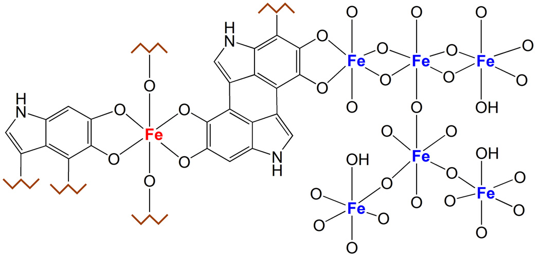
Fig. 4. Iron centers in NM pigment.
The figure shows the two types of iron centers present in the NM pigment. The multinuclear iron cluster, similarly to ferritin, contains iron(III) ions (blue colored in the figure) coupled by oxy-hydroxy bridges and surrounded by catechol groups of NM. In this site, iron is probably stored with high affinity and maintained in a redox inactive state, and is principally detected by Mössbauer spectroscopy. The other iron site consists of mononuclear centers, where iron (red colored) is coordinated by oxygen atoms of catechols moieties, and possibly by hydroxo groups. This could be a low affinity binding site occupied only in case of iron overload, when the high affinity centers are saturated; in iron overload condition as occurring in PD, the mononuclear iron could be still redox reactive and catalyze the production of toxic species, for example via the Fenton’s reaction. Iron in this site is principally detected by EPR spectroscopy.

