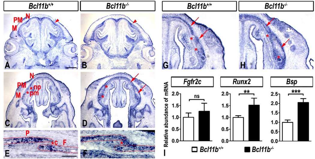
Fig. 6. Altered Fgfr2c expression in the Bcl11b−/− embryonic faces and coronal suture.
(A–H) RNA ISH using an Fgfr2c probe in sections of embryonic faces at E14.5 at two different planes (A–D) and at higher magnification (G, H) and in coronal suture at E16.5 (E, F). Arrowheads denote up-regulation and expansion of Fgfr2c expression. Arrows point to the lack of Fgfr2c expression within the central osteoid of facial bones in Bcl11b−/− mice. Asterisks indicate ectopic expression of Fgfr2c in Bcl11b−/− sutures. Bones: F, frontal; M, maxillary; N, nasal; P, parietal; PM, premaxillary. Sutures: c, coronal; np, naso-premaxillary; pm, premaxillary-maxillary. Scale bars: (A–D) 500 µm; (E, F) 200 µm; (G, J) 100 µm. (I) qRT-PCR analysis indicates quantitatively non-significant change in Fgfr2c expression and an increase in Runx2 and Bsp expression in Bcl11bncc−/− faces at E14.5. Error bars indicate standard deviation. ns, not significant; ** denotes statistical significance at p ≤ 0.01; *** denotes statistical significance at p ≤ 0.001 (n = 3).

