
Fig. 3.
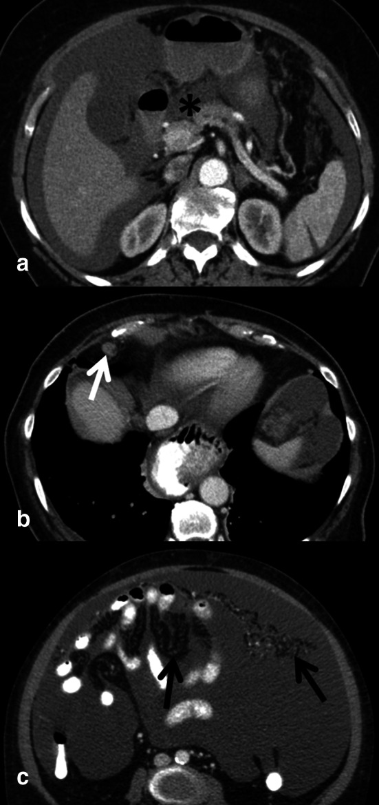
Subtle imaging findings indicative of advanced ovarian cancer spread: ascites in omental bursa (a, asterisk) lymph node (b, arrow) with a short axis diameter of >7 mm in the cardiophrenic fat above the diaphragm. In all ovarian cancer staging exams, mesentery and omentum should be scrutinized for band-like and reticular pattern (c, arrows) presenting peritoneal spread