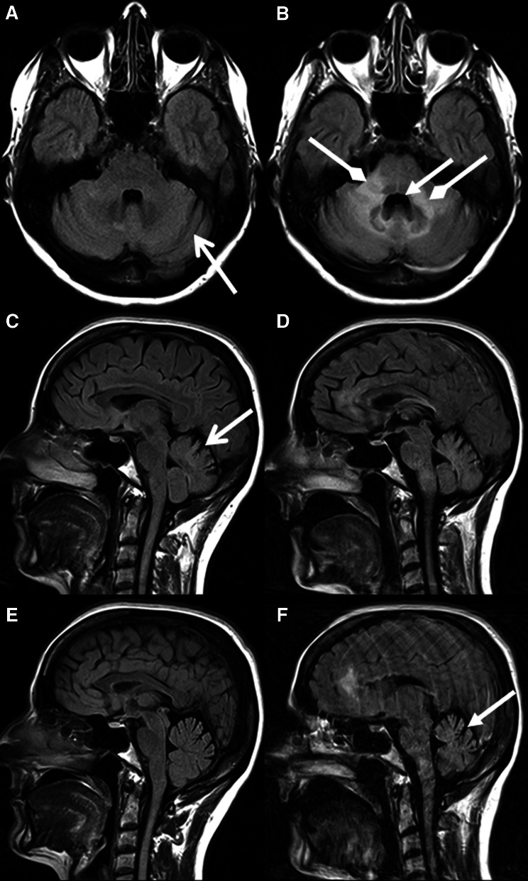
Fig. 3.
Axial fluid attenuated inversion recovery (FLAIR) images (a, d), and sagittal FLAIR images (b, c, e, f) of patient 3 showing grade 1 cerebellar atrophy at the time of PML diagnosis (a–c) and grade 2 cerebellar atrophy 3 months later (d–f). There were already bilateral dilated sulci at diagnosis (open arrowhead). However, there was clear loss of cerebellar volume 3 months later. Again, note the enlarged fourth ventricle (closed arrowheads). In addition, large hyperintense lesions in the cerebellar peduncles and brainstem developed (diamond arrowheads)

