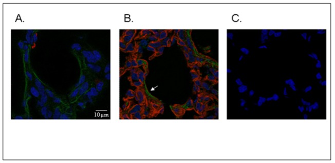
Figure 3. SPOCK2 immunofluorescence studies in rat lung tissue on postnatal day 14.

Immunocolocalization of SPOCK2 (green labelling) and SP-B (red labelling, A) or collagen IV (red labelling, B) showed the presence of SPOCK2 throughout the ECM, including the basement membrane, as shown by superimposing the 2 fluorescent signals in some areas (B, white arrow). Negative control lung incubated with mouse and rabbit polyclonal IgG, showing fluorescence restricted to the nucleus (C.) (original magnification x126).
