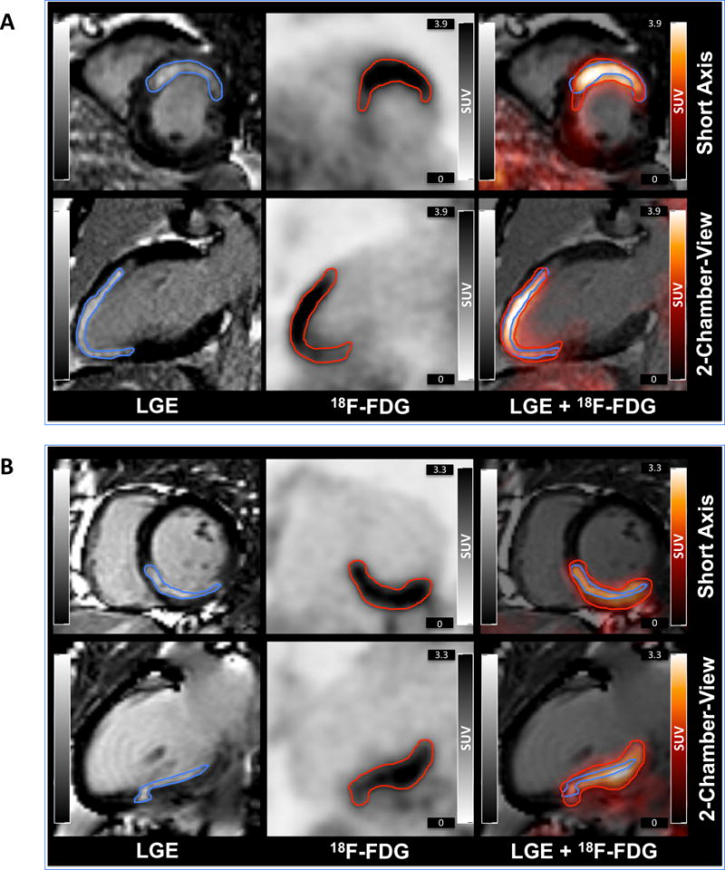
Figure 2. 18F-FDG PET/MR images of two patients shortly after AMI.

Short and long axis views of LGE MR images (left), 18F-FDG PET images (middle), and overlay (right) of patients with anterior (A) or inferior (B) myocardial infarction.

Short and long axis views of LGE MR images (left), 18F-FDG PET images (middle), and overlay (right) of patients with anterior (A) or inferior (B) myocardial infarction.