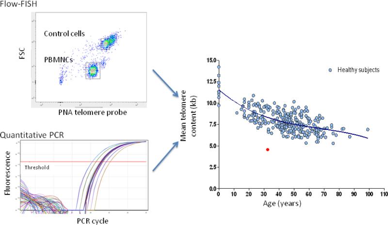
Figure 4.
Telomere content of leukocytes measured by standardized flow FISH or quantitative PCR. A patient's result is compared to age-adjusted normalized values. Calculated telomere length of total mononuclear cells below the first percentile for age strongly suggests a diagnosis of telomere disease. (Courtesy of Dr. Bogdan Dumitriu)

