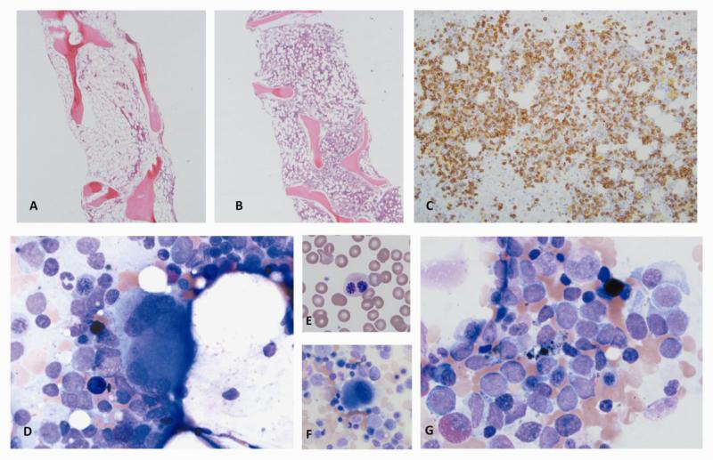
Figure 5.
GATA2 deficiency in the clinic: The patient presented as 18 year-old male with pancytopenia and marrow of AA, and within two years, he developed AML with myelodysplastic morphology.
A. Initial bone marrow biopsy at presentation: hypocellular marrow with trilineage hypoplasia compatible with AA. B-E. Bone marrow biopsy two years later: B. AML with myelodysplastic morphology; 30-40% cellularity. C. CD34 immunohistochemistry of biopsy, highlighting increased blasts. D. Dysplastic large osteoclast-like megakaryocyte with separated nuclear lobes, on aspirate smear. E. Pelgeroid PMN, peripheral smear. F. Small mononuclear megakaryocyte, on aspirate smear. G. Increased blasts, 35% on 500 cell differential of aspirate smear. (Courtesy of Dr. Danielle Townsley and Dr. Katherine Calvo).

