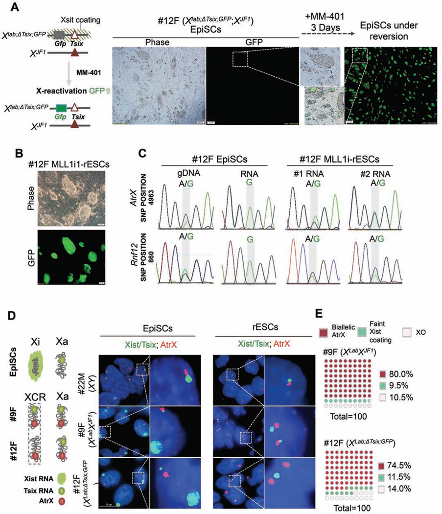
Figure 2. Reactivation of Xi-chromosome during EpiSC reversion.
A. Left, X-chromosome allele information for 12F female EpiSCs. Right, representative phase/ GFP images of 12F EpiSCs before and after MM-401 treatment. B. Representative images of phase/GFP for Mll1i-rESC after passages 2. C. SNP sequencing for Atrx and Rnf12. The divergent nucleotides in SNP position were highlighted in grey. D. Left, schematics for X-chromosome status in EpiSCs and ESCs. Right, RNA-FISH for Xist/Tsix (green) and Atrx (red) in EpiSCs and Mll1irESCs. Scale bar, 10µm. E. Quantification of biallelic expression of Atrx (red) or incomplete Xi reactivation in Mll1i-rESCs. XO, cells with only one detected X-chromosome.100 nuclei were counted for each cell line from two independent experiments. This Figure is related to Figure S3.

