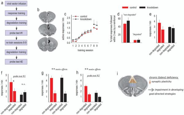
Figure 3. Adolescent-onset GABAAα1 silencing impairs goal-directed action selection in adulthood.
(a) Experimental timeline. (b) Histological representations of infusion sites are transposed onto images from the Mouse Brain Library (Rosen et al., 2000). Black indicates the largest regions affected, and white the smallest. Infusions were bilateral. (c) Mice were trained to respond for food reinforcers; breaks in the acquisition curves annotate tests for sensitivity to action-outcome contingency degradation. Response rates represent both trained responses/min. (d) Following training, the schedules of reinforcement were modified such that roughly half of responses directed towards one nose poke recess were reinforced, while pellet delivery followed only ~7% of responses directed towards a different nose poke recess. These contingencies are referred to as “non-degraded” and “degraded.” (e) Response rates during these training sessions are shown. (f) During a subsequent probe test, control mice preferentially generated the response most likely to be reinforced in a goal-directed manner (“non-degraded”). Mice with Gabra1 knockdown instead generated both responses at equivalent rates, a failure in action-outcome conditioning. (g) With additional task experience, both groups generated higher response rates during training sessions when the response was likely to be reinforced. (h) Correspondingly, mice were also ultimately able to select actions that were more, vs. less, likely to be reinforced in a probe test. (i) Our model: Chronic Gabra1 deficiency has been associated with decreased synaptic marker expression and decreased expression of mushroom-shaped spines in the cortex (see Discussion). We suggest that these abnormalities in the prelimbic cortex (highlighted at center) delay the development of goal-directed response strategies. The anterior cingulate (dorsal) and the medial orbitofrontal cortex (ventral) are also highlighted on this image from the Mouse Brain Library (Rosen et al., 2000). Means+SEMs,*p<0.05,**p≤0.01. n=5/group.

