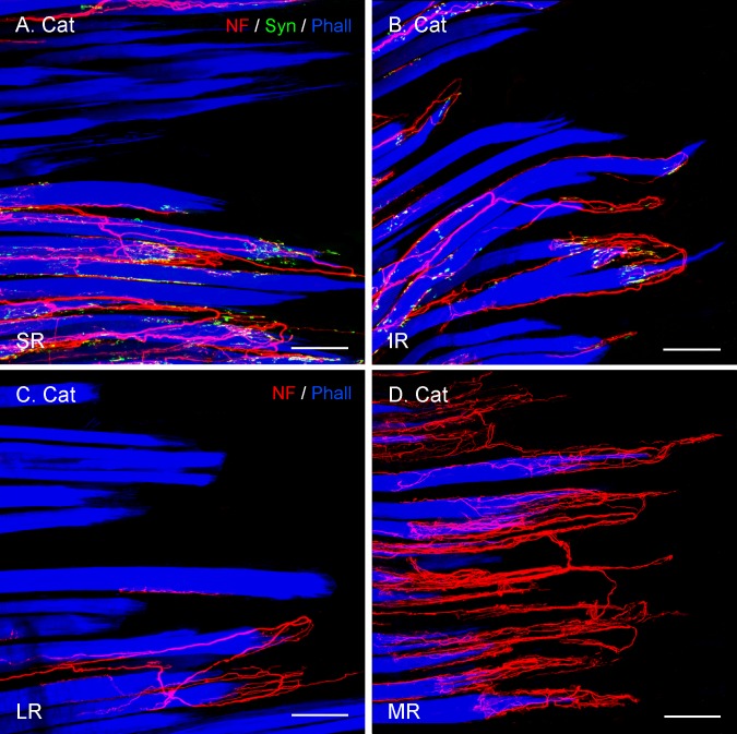
Figure 3.
Projection of CLSM z-stacks showing segments of the distal muscle–tendon junction of the four rectus muscles (SR, IR, LR, MR) in cat. (A, B) Axons were labeled with anti-neurofilament (NF, red), anti-synaptophysin (Syn, green), and muscle fibers with phalloidin (Phall, blue). (C, D) Staining with anti-neurofilament and phalloidin, but lacking anti-synaptophysin labeling. (A–D) The relative EOM-specific abundance of palisade endings with the medial rectus (D) highest and the lateral rectus (C) lowest. The values of the vertical eye muscles (A, B) are in-between. Scale bars: 100 μm.

