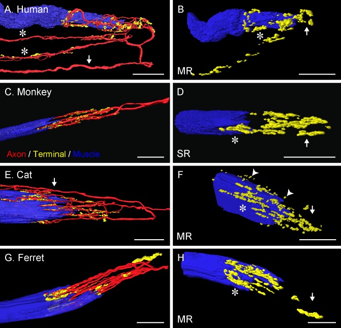
Figure 4.
Three-dimensional reconstruction of palisade endings in frontal-eyed species. In monkey (C), the palisade ending is from an SR muscle; in the other species they are from MR muscles. Nerve fibers were labeled with anti-neurofilament (red), terminal varicosities with anti-synaptophysin (green), and muscle fibers with phalloidin (blue). The yellow color represents the merging of green (synaptophysin) and red (neurofilament) signals. (A, E) Showing the whole formation of the palisade ending with the axon (arrows) indicated. (A) The axon supplies three palisade endings, of which only the upper one is completely shown. Asterisks indicate the collateral supplying the other palisade endings. (C, G) From the axon supplying the palisade endings, only the recurrent part coming from the tendon is shown. (B, D, F, H) Showing the same palisade endings as in (A, C, E, G), respectively, but rotated and the nerve fibers removed. Terminal varicosities are found far away from the muscle fiber (arrows), at the tendon level, and around the muscle fiber tip (asterisks). Many nerve terminals around the muscle fiber tip are separated from the muscle fiber surface by a small gap (arrowheads [F]). Scale bars: 30 μm.

