. 2016 Mar;18(1):99–107. doi: 10.31887/DCNS.2016.18.1/kparker

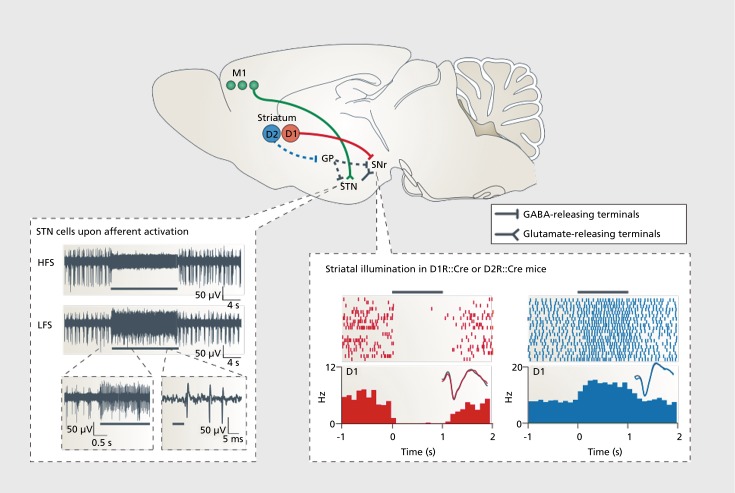
Figure 4. A schematic diagram (top panel) shows key neural projections that are involved in parkinsonian behavior and treatment. Data in the bottom left panel are from a study that used a constitutively expressing ChR2 mouse line (Thy1::ChR2) to identify a mechanistic explanation for the therapeutic effects of deep brain stimulation. By illuminating and recording in the subthalamic nucleus, this paper showed that afferent fibers entering the subthalamic nucleus, rather than local cell bodies themselves, are likely to be the direct target of deep brain stimulation in the correction of parkinsonian motor activity. High-frequency stimulation of the afferent fibers into subthalamic nucleus potently silenced the structure as shown and reversibly abolished the parkinsonian symptoms. By contrast, low-frequency stimulation of the afferents simply added spikes on top of endogenous spikes and worsened parkinsonian symptoms.12 Data in the bottom right panel are from a study that used a Cre-AAV to selectively express ChR2 in either D1 dopamine receptor (D1R)::Cre or D2 dopamine receptor (D2R)::Cre mice to examine the differential contributions of the direct and indirect pathways with respect to motor output. Activation of D1R-expressing neurons silenced local basal ganglia activity and increased ambulation, whereas activation of D2R-expressing neurons increased this activity and enhanced immobile or bradykinetic (slow) behavior.45 Black bars indicate the duration of illumination. AAV, adeno-associated viral; ChR2, Channelrhodopsin-2; HFS, high-frequency stimulation; LFS, low-frequency stimulation; M1, primary motor cortex; μV, microvolts; D1, D1-type dopamine receptor; D2, D2-type dopamine receptor; GABA, γ-aminobutyric acid; GP, globus pallidus; Hz, hertz; ms, milliseconds; s, seconds; SNr, substantia nigra pars reticulata; STN, subthalamic nuclus. Reproduced from reference 19: Tye KM, Deisseroth K. Optogenetic investigation of neural circuits underlying brain disease in animal models. Nat Rev Neurosci. 2012;13(4):251-266. © 2012, Nature Publishing Group. Bottom left image group originally published in reference 12: Gradinaru V, Mogri M, Thompson KR, Henderson JM, Deisseroth K. Optical deconstruction of parkinsonian neural circuitry. Science. 2009;324(5925):354-359. © 2009, American Association for the Advancement of Science. Bottom right image group originally published in reference 45: Kravitz AV, Freeze BS, Parker PRL, et al. Regulation of parkinsonian motor behaviours by optogenetic control of basal ganglia circuitry. Nature. 2010; 466(7306): 622-626. Copyright © Nature Publishing Group, 2010.

Figure 4.