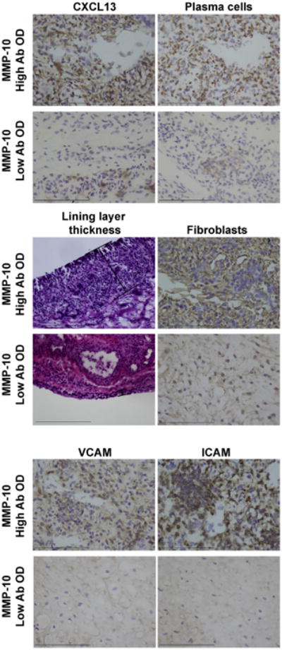
Fig. 10.

Immunohistochemical staining of synovial tissue from two representative patients with antibiotic-refractory LA who had high or low rankings for serum MMP-10 IgG antibodies. The patient with the highest ranking for MMP-10 antibodies had marked staining for CXCL13, plasma cells, lining layer thickness, fibroblast proliferation, and expression of the cellular activation markers VCAM and ICAM. In contrast, the patient with the lowest ranking had minimal staining for these histologic findings. Brown indicates specific staining of the immune cells and purple is the counter stain (hematoxylin). Lining layer thickness was assessed by hematoxylin and eosin (H&E) stain. Bars = 100 μm. (For interpretation of the references to color in this figure legend, the reader is referred to the web version of this article.)
