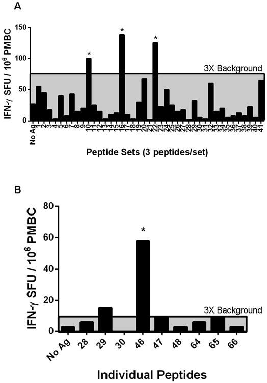
Fig. 2.

T cell autoreactivity with peptides isolated from synovial tissue of patient LA4. Screening of 124 HLA-DR-presented peptides identified from the synovial tissue of one patient (LA4) for T cell reactivity using the patient's own PBMC, as measured with the human IFN-γ ELISpot assay. A. Peptides isolated from the synovial tissue of patient LA4 were synthesized and tested in pools with the patient's PMBC. A positive result was defined as >3× background of unstimulated cells (area above the gray shaded region). Stars indicate the 3 peptide pools that were >3× background. B. Individual peptides from the 3 positive peptide pools were retested with PBMC from patient LA4. A positive result was defined as >3× background of untreated cells (area above the gray shaded region). The star indicates the peptide with the greatest reactivity, which was derived from the source protein MMP-10.
