Fig. 6.

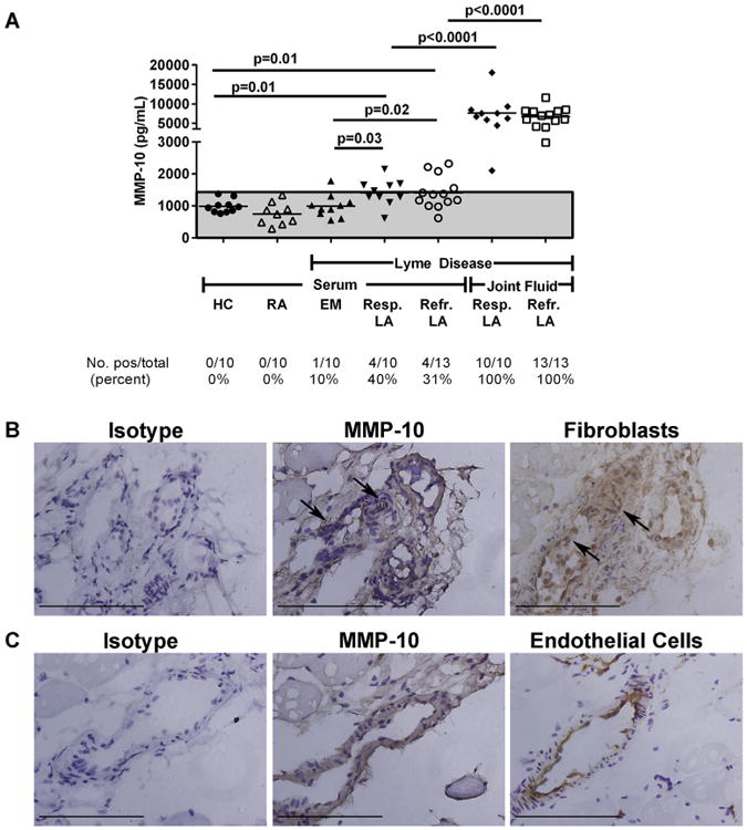
MMP-10 protein in Lyme disease patients and control groups. A. MMP-10 protein concentrations were measured in matched serum and synovial fluid samples in patients with antibiotic-responsive or antibiotic-refractory Lyme arthritis (LA), and in serum samples from patients with erythema migrans (EM) or rheumatoid arthritis (RA), and in healthy controls (HC) subjects. MMP-10 protein concentrations are shown, as measured by Luminex assay. The area above the shaded gray area is >3 standard deviations above the mean value of HC. P-values were calculated using Mann–Whitney test. (B + C) The panels in parts B and C are serial sections from the same area of the slides. Bars = 100 μm. B. MMP-10 staining is observed in the synovial tissue and is associated with staining for synovial fibroblasts (arrows). No staining is observed in the mouse isotype control. C. MMP-10 staining is observed in the synovial tissue and is associated with staining for endothelial cells. No staining is observed with the mouse isotype control.
