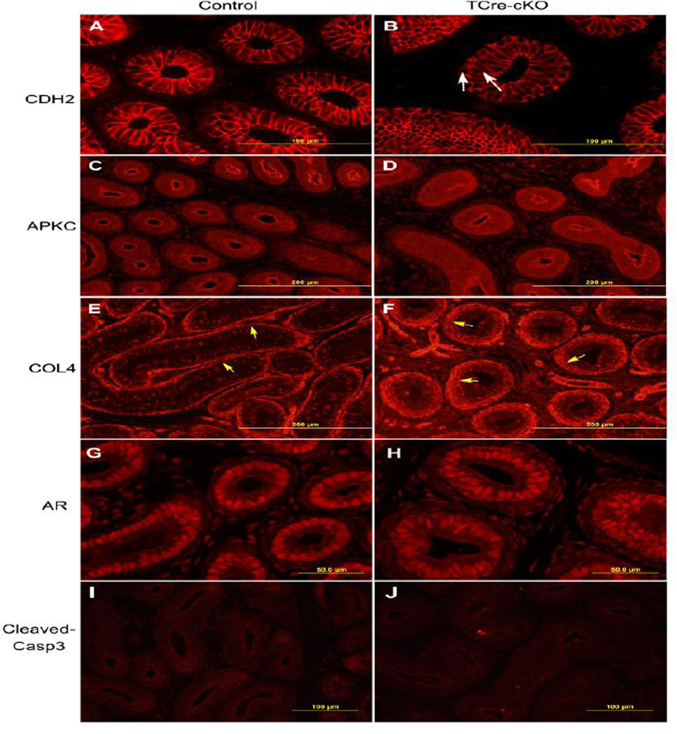
Fig. 3.
Identification of adhesion junctions, apical-basal polarity, basement membranes, AR localization, and apoptotic cells in the control and TCre-cKO epithelium at P14. (A–B) Representative images of localization of adhesion junction component CDH2 in controls and TCre-cKOs. White arrows show pseudostratified epithelial cells. (C–D) Representative images of apical localization of atypical PKC (APKC) in controls and TCre-cKOs. (E–F) Representative images of COL4 localization in controls and TCre-cKOs. Yellow arrows show the basement membrane. (G–H) Representative images of AR localization in controls and TCre-cKOs. (I–J) Representative images of cleaved caspase3 localization in controls and TCre-cKOs.

