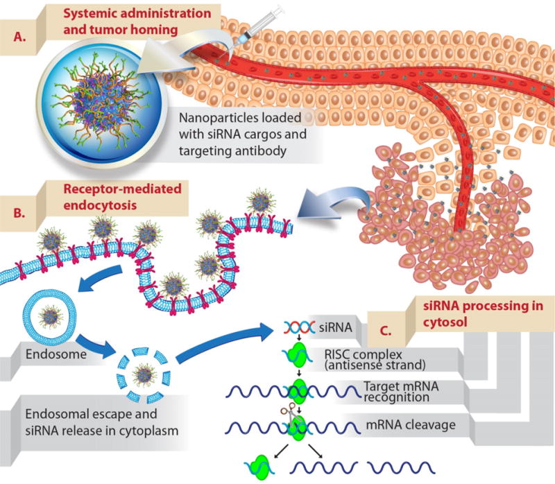
Figure 2. Schematic illustration of delivering siRNAs to tumors upon intravenous administration.

(A) Nanoparticles (mesoporous silica nanoparticles surface functionalized with polymer and targeting antibody used as an example) that were developed as siRNA carriers need to be able to protect siRNAs from blood degradation and prolong their blood residence time. This will enhance the propensity of siRNA-nanoparticles to accumulate in tumors via the EPR effect (see text). (B) Nanoparticles are typically taken up to cancer cells by endocytosis. The uptake can be further promoted by conjugating targeting ligands onto nanoparticles (active targeting). Upon endocytosis, siRNAs need to escape endosome to their site of action, cytosol. (C) Delivered siRNAs are then processed by intracellular machinery (RNA-induced silencing complex) and degrade their target complementary mRNA.
