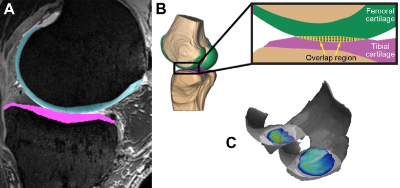
Figure 5.
(A) Manually segmented cartilage from magnetic resonance imaging. (B) A nondeformable cartilage model is created and coregistered with computed tomography and dynamic stereo x-ray testing data. The model’s overlap region is calculated, which allows determination of contact location, area, and strain. (C) Example of evaluation of the overlapped region for 1 specimen.

