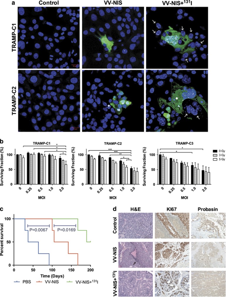
Figure 7.
Efficacy of GLV-1h153 in TRAMP models. (a) Confocal images of H2Ax foci resulting from DNA double-strand breaks in TRAMP cells treated with GLV-1h153 and 131I. Blue: DAPI, Green: Viral GFP, White: γH2Ax foci. White arrows mark the non-infected ‘bystander' cells that have received DNA damage. (b) TRAMP cells treated with GLV-1h153 and external beam radiation. Reduction of proliferative capability was measured by MTT assay at 48 h post infection. s.e.m.s are shown. Significance is the result of two-way ANOVA with Bonferroni multiple comparisons test, *P<0.05, **P<0.001, ***P<0.0001. (c) Long-term survival of TRAMP mice bearing spontaneous prostate tumors and treated intravenously with 5 × 10⁷ PFU GLV-1h153 (VV-NIS) and 1 mCi 131I (VV-NIS+131I) (n=4). Kaplan–Meier plot significance is the result of log-rank (Mantel Cox) test. (d) Representative examples of the histology of the above TRAMP mouse prostates, showing H&E staining and IHC for KI67 and probasin at the time of death.

