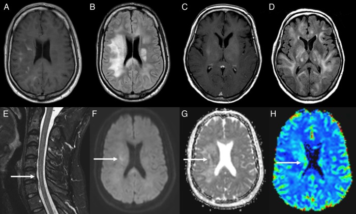
Fig. 1.
(A and B) MRI of a young male (patient 40) showing patchy contrast enhancement in the right hemisphere in axial T1 sequence (A) and diffuse hyperintense lesions in bilateral white matter on FSE-FLAIR sequence (B). (C and D) MRI of an adult female (patient 36) showing contrast in right subinsula and left thalamus (C) and diffuse bilateral abnormal hypersignal within the deep white matter in FLAIR sequence (D). (E–H) Short-TI inversion-recovery (STIR) spinal cord MRI from patient 40 showing an intramedullary hyperintense lesion (arrow) (E). Diffusion-weighted image shows elevated signal in right hemisphere (arrow) (F), and ADC map (G) shows (arrow) low signal in the lesion confirming that it is a true restricted diffusion. Perfusion-weighted image shows an elevated relative regional cerebral blood volume ratio (H).

